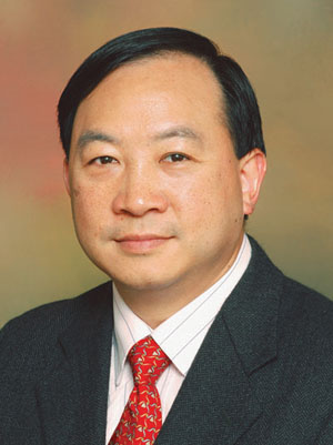
Prof. Cheung Lim-kwong was the Associate Dean of Research, the former Chairman of the Faculty Higher Degrees Committee at the HKU Faculty of Dentistry and a member of the Board of Graduate Studies. Prof. Cheung's PhD thesis titled The temporalis myofascial flap in maxillofacial reconstruction: vascular anatomy and healing was previously available for downloading at HKU Scholars hub at http://hub.hku.hk/bitstream/10722/128702/1/FullText.pdf . However following the exposure of the plagiarism scandal HKU has blocked online access to this thesis.
A side by side comparison reveals the degree of plagiarism. This may be only the tip of the iceberg. Most journal articles during the 1990's & 80's are only available in the hard copy format. Prof. Cheung has also included in the PhD, paragraphs copied from some text books. So it is quite difficult to locate other potential sources he may have plagiarized.
Page No.
|
Prof. Lim Cheung's PhD
|
Original Text
|
Original Source
|
5
|
The temporalis muscle inserts into the coronoid process and the anterior aspect of the ramus of the mandible.
|
The muscle inserts into the coronoid process and anterior aspect of the vertical ramus of the mandible.
|
|
7
|
The superficial temporal fascia is a thin, highly vascular layer of moderately dense connective tissue. It lies immediately deep to the hair follicles and the subdermal layer of fibrofatty tissue.
|
The temporoparietal fascia is a thin layer of moderately dense connective tissue, which lies immediately deep to the hair follicles and subdermal fibro-fatty tissue.
| |
7
|
.... is part of the superficial musculoaponeurotic system and is continuous with the fascia over the
parotid gland below and with the galea aponeurosis above.
|
is part of the subcutaneous musculo-aponeurotic system, and is continuous with the galea superiorly and the superficial musculo-aponeurotic system inferiorly
| |
7
|
The superficial temporal fascia is separated from the deep temporal fascia by a distinct layer of loose areolar tissue.
|
It is separated from the underlying deep temporal fascia and temporalis muscle by a loose avascular plane.
| |
7
|
The deep temporal fascia is a dense, uniform fascial layer which completely invests the outer aspect of the temporalis muscle. At the periphery of the muscle all round, the fascia fuses with the pericranium at the superior temporal line.
|
.....the deep temporal fascia. This is a dense fascial layer which completely invests the outer surface of the muscle from the upper edge of the zygoma, and peripherally fuses with pericranium at the superior temporal line of the skull.
| |
10
|
obliterating the dead space following orbital exenteration (Naquin 1956, Webster 1957, Reese 1958, Reese & Jones 1961), and
|
obliteration of dead space following orbital exenteration (Naquin, 1956; Reese and Jones, 1961; Deitch and
| |
18
|
Considerable interest has developed in the combined use of the temporalis muscle and its underlying periosteum to provide a single vascularized unit with bone forming capacity.
|
Most recently, considerable interest has developed in the combined use of temporalis muscle and the underlying pericranium to provide a single vascularised unit with bone-forming capacity.
| |
18
|
The osteogenic potential of the temporal musculoperiosteal flaps has been confirmed experimentally in young animals (Hauben & van der Meulen 1984).
|
The osteogenic potential of temporal musculoperiosteal flaps has been confirmed experimentally in young animals (Hauben and van der Meulen, 1984)
| |
18
|
Its immediate clinical application, however, is restricted by the possible deleterious effects that early masticatory muscle transposition may have on subsequent craniofacial growth (Hohl 1983).
|
Its immediate clinical application is, however, restricted by the possible deleterious effects that early masticatory muscle transposition may have on subsequent craniofacial growth (Hohl, 1983).
| |
72
|
..the anterior and posterior deep temporal vessels supply the anterior and posterior part of the muscle respectively...
|
...the anterior and posterior deep temporal arteries, supply the anterior and posterior portions of the muscle, respectively
(Mathes and Nahai, 1982).
| |
9
|
Lexer (1908) and Rosenthal (1916) utilized the TMF to reanimate the eyelid following paralysis of the facial nerve.
|
Lexer (1908) and Rosenthal (1916) utilized the TMF to reanimate the eyelid following paralysis of the facial nerve,
|
|
11
|
Ewers (1988) described a different technique of reconstruction after maxillectomy with the.....
|
Ewers (1988), reported a new technique of reconstruction of the
| |
12
|
Colmenero et al. (1991) reported their experience of 26 TMFs....
|
Colmenero et al. (1991), reported their experience gained with 26 temporalis flaps....
| |
12
|
...as a composite flap by combining it with cranial bone, coronoid process or a temporal skin island. Although major complications were not observed,
|
.....as a composite flap with cranial bone, coronoid process or skin island. Major complications were not observed.
| |
12
|
total necrosis of the flap developed in 3 cases
|
,total necrosis of the TMF occurred in 3 cases
| |
12 & 13
|
should be taken into consideration before deciding on more extensive reconstructive procedures.
|
should be taken into consideration before deciding on more extensive reconstructive procedures.
| |
13
|
Van der Wal & Mulder (1992) reported another 4 cases of large palatal defects in cleft lip and palate
|
Van der Wal and Mulder (1992), reported closure of large palatal defects in four patients with congenital cleft palate.
| |
38
|
the residual posterior part of the muscle transposed to fill the anterior temporal fossa,
|
The posterior part of the muscle was then advanced to fill in the depression in the temporalis fossa.
| |
9
|
The first use of the temporalis myofascial flap (TMF) has been attributed to Golovine
|
the origin of the temporalis muscle flap which has been attributed to Golovine,
|
|
9
|
Sir Harold Gillies in 1917 described a series of cases where the temporalis muscle was used as a transposition flap for deformities caused by the loss of the zygomatic bone,
|
Sir Harold Gillies in 1917 described a series of cases where temporalis muscle was used as a transposition flap for deformities caused by loss of the malar bone.
| |
9
|
In a later paper (Gillies 1934), he described the use of
TMF, tunnelled to
|
In a later paper (Gillics, 1934) he described the use of temporalis muscle and fascia tunnelled to
| |
9
|
...either the corner of the mouth or the inner canthus of the eye, for facial reanimation.
|
...either the corner of the mouth or the inner canthus of the eye for reanimation.
| |
5
|
The temporalis muscle inserts into the coronoid process and the anterior aspect of the ramus of the mandible.
|
...the temporalis msucle is inserted into the coronoid process and the anterior border of the ramus of the mandible...
| |
5
|
DuBrul (1980) pointed out that the tendinous attachment to the mandible can be divided into the
superficial and deep tendons. The superficial tendon inserts into the anterior border of the coronoid process and the deep tendon inserts into the internal oblique line reaching down to the retromolar pad.
|
DuBrul (1980) presents further detail stating that there are superficial and deep tendons which the former inserts into the anterior border of the coronoid process and the latter inserts into the temporal crest (internal oblique line) reaching the area of the lower third molar into the retromolar pad.
| |
54
|
The fact that glycogen decreases as the degree of keratosis increases suggests a role for glycogen in keratinization. The glycogen may serve as a source of energy for keratin synthesis.
|
The fact that glycogen content decreases as the degree of keratosis increases suggests a role for glycogen in keratinization. The glycogen may serve as a source of energy required for keratin synthesis
|
|
92
|
.....with the marginal cells at the advancing front being the active motile cells, while the cells behind the margins are passively dragged along
|
the cells at the margin of the moving sheet appeared to be actively motile while the cells behind (or above, in a stratified layer) the marginal cells were passively dragged along
| |
92
|
This mode of sheet movement, referred to as the sliding model of wound closure, has been demonstrated on epithelial cells in cell culture (Vaughan & Trinkaus 1966), in embryo (Bellairs 1963), and in corneal healing (Buck 1979).
|
This mode of sheet movement, referred to as the sliding model of wound closure, has
been demonstrated directly for epithelial cells
in tissue culture,36 for embryonic epithelial
moveme nt,38 for amphibian wound closure,37
and for corneal wound closure.39
|
Page 119.
|
92 & 93
|
It is more difficult to study mammalian cutaneous wound closure directly because of the thickness and opacity of the dermis. Moreover, the migrating epithelial sheet in the mammalian epidermis is multilayered and thus more complex than those systems illustrated in the sliding model.
|
It is much more difficult to study mammalian
cutaneous wound closure directly because of the thickness and opacity of the dermis. Moreover, the migrating epithelial sheet of mammalian epidermis is multilayered and thus more complex than those systems illustrating the sliding model.
| |
93
|
For the repair of mammalian epidermis, Winter (1964) proposed the "leap frog model" of epidermal sheet movement. This model was deduced from morphological data at ultrastructural level, which suggested that cells at the migrating front adhere to the substrate......
|
For the repairing mammalian epidermis, Winter40 proposed the leap frog model of epidermal sheet movement (Fig. 7- 4). This model was deduced indirectly from ultrastructural morphological data that suggested that cells at the migrating front adhere to the substrate.........
| |
93
|
.......submarginal cells are thus conceived to crawl over the newly adherent basal cells in a "leap frog" fashion (Krawczyk 1971, Kuwabara et al. 1976, Gibbins 1978).
|
.......submarginal cells are conceived to crawl over the newly adherent basal cells in a "leapfrog" fashion
| |
93
|
What actually turns on the cellular machinery of movement, be it physical or chemical, is still not known.
|
what actually "turns on" the cellular machinery of movement, be it physical or chemical, is still not known.
| |
93
|
Little work has been done on the cytoskeletal mechanism of epidermal cell motility, but it is recognised that epidermal cells in all strata of the
epidermis contain actin and the motile machinery probably involves the actin-myosin contractile system
|
Little work has been done on the cytoskeletal mechanisms of epidermal cell motility, but it is recognized that epidermal cells in all strata of the epidermis contain actin and that the motile machinery probably involves the actin-myosin contractile system.
| |
93
|
A cytoskeletal model of epidermal cell motility has been proposed by Bereiter-Hahn et al. (1981), in which the motive force is generated by directed contractions of the actin filaments in the cell body, forcing the cytoplasm .....
|
A cytoskeletal model of epidermal cell motility has been proposed by Bereiter-Hahn and associates45 in which the motive force is generated by directed contractions
of the actomyosin system, forcing cytoplasm
....
| |
93
|
It is generally held that a free edge is all epithelium needs to initiate movement....
|
It is generally held that a free edge is all epithelium needs to initiate movement.
| |
93
|
However, this concept may be an oversimplification since epidermal cells will not migrate in cell culture unless the substratum is optimal even though the epidermal cells have a free edge...
|
However, that concept may be too simple since primary epidermal cells not adapted to culture will not spread in tissue culture unless the substratum is optimal even though the cells have a free edge......
| |
93
|
....it appears likely that a free edge may provide the stimulus for the epithelial cells to spread, but .....
|
....it appears likely that a free edge may provide the external stimulus to spread, but ....
| |
84
|
Recent experiments support the fact that under certain circumstances, mesenchymal cells may transform to become part of the regenerating
epithelium (Chong et al. 1987).
|
Recent observations suggest, however, that under some circumstances mesenchymal cells may transform and become part of the regenerating epithelium;26
|

No comments:
Post a Comment